
Disease control and Management
Back
Disease control and Management
• Managing disease risk is an important component for livestock operations. Whether it is part of everyday activities or in the event of an outbreak, awareness of common biosecurity, or biological risk management, protocols are essential to those interacting with animal facilities.
• Managing disease risk must be done on a daily basis to ensure the health of livestock and livestock producers.
• However, there are specific prevention steps for certain diseases, and control practices relate to how the disease is actually spread.
• The five routes of transmission will be further explained but include aerosol, direct contact, fomites, oral and vectors. There are commonalities in disease prevention, but some diseases will require additional specific responses to prevent and control spread.
• Heightened biosecurity will be needed during an animal health event and afterwards to minimize or eliminate disease spread.
Infectious Diseases of Cattle and Buffalo
Infectious diseases, also known as communicable diseases, contagious diseases or transmissible diseases comprise clinically evident illness (i.e., characteristic clinical signs and/or symptoms of disease) resulting from the infection, presence and growth of pathogenic biological agents in an individual host organism. In certain cases, infectious diseases may be asymptomatic for much or all of their course. Infectious pathogens include some viruses, bacteria, fungi, protozoa, multicellular parasites, and aberrant proteins known as prions. These pathogens are the cause of disease epidemics, in the sense that without the pathogen, no infectious epidemic occurs.
Transmission of pathogen can occur in various ways including physical contact, contaminated food, body fluids, objects, airborne inhalation, or through vector organisms. Infectious diseases that are especially infective are sometimes called contagious and can be easily transmitted by contact with an ill person or their secretions. Infectious diseases with more specialized routes of infection, such as vector transmission or sexual transmission, are usually regarded as contagious but do not require medical quarantine of victims.
Mastitis
About the Diseases
Nature of disease
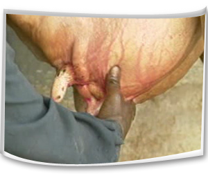
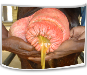
• Mastitis is the inflammatory condition of the udder irrespective of causes.
• It manifests the changes in the milk colour and consistency.
• Milk yield reduces abruptly and results in heavy economic loss.
• High yielding dairy cows are more commonly affected than low yielders.
• Exotic and cross bred cows are more prone to mastitis than the Indian zebu cows.
Causes
• A large number of species of microorganisms have been implicated as causes of mastitis. They are bacteria, fungus, Mycoplasma and virus.
• The most important bacterial organisms causing mastitis areStaphylococcus aureus; Str. agalactiae; Str.zooepidemicus; Str.faecalis; Str. pyogenes; Klebsiella spp; Mycobacterium bovis; E.coli; Brucella abortus; Pseudomonas pyocyaneus; Leptospira pomona; Pasteurella multocida.
• The fungal organisms responsible for mastitis are Trichosporon spp; Aspergillus fumigatus; A.midulus; Candida spp;
• Hygiene, trauma, complete milking and teat injuries may predispose this condition.
Mode of Transmission
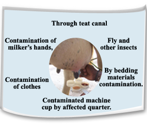
• Through the teat canal infection reaches the mammary gland.
• The normal inhabitant of udder and environment like Str. Agalactiae,Stap.aureus, E.coli and Ps.pyocyaneus under favourable conditions multiply and invade the tissues produce much damaging effect.
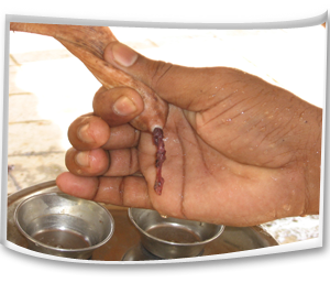
• The cutaneous surface of the cow may have many organisms as resident population and from where the organisms may have the chance of invade through contamination by handlers.
• The contamination of milker’s hands, clothes and machine cup by milk from the affected quarter may lead to the spread of the disease to other non-infected teats of cow.
• Fly and other insects may also spread the infection from one place to the other.
• Spread of infection is possible through bedding ground by discharges of affected gland.
Symptoms
Clinical symptoms
• Swelling of udder as a hard mass.
• Swollen udder with hot and pain while touching it.
• Animal will not allow touching the udder and will kick while touching it.
• Swollen and reddening of teats.
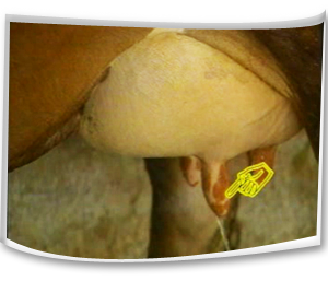
• Milk mixed with blood.
• Milk mixed with yellow or brown fluid with flakes or clots with foul smelling.
• Reduced milk yield.
Management Methods
Preventive measures
• Cow should be allowed in soft bedding following parturition.
• Concrete floor should be avoided especially in case of high yielder. Bedding should be done with straw, saw dust or sand. Sand is the ideal bedding material since it has lower bacterial count.
• Infusion should be used in each cow at dried off.
• Always the animal sheds should be clean.
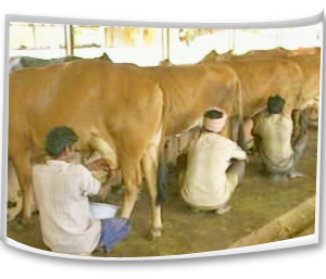
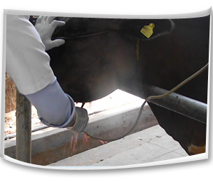
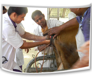
• Washing the udder and hand of the milker with antiseptic lotion (4% Pottasium permanganate solution) before and after milking.
• The floor of the milking shed should be washed with running water.
• The milker’s hand should be free from nail.
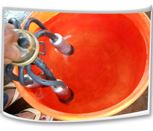
• Cleaning and disinfecting milking machine and the teat cup, vessels after each milking.
• The healthy non-infected cows should be milked first and known infected cows should be milked at last.
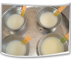
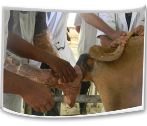
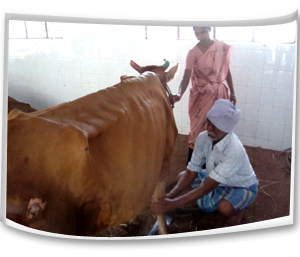
• Newly introduced cow should be milked separately and should be screened through California Mastitis Test (CMT).
• The first strip of milk should not be allowed to fall on the floor; they may be stripped in separate container along with disinfectants in it.
• Dipping of all teats following each milking with iodophor solution containing 1% available iodine or hypochlorite solution and Chlorhexidine in 0.5% to 1% polyvenylpyrrolidine solution.
• Immediately after milking should not allow the animal to lie-down by engaging with fodder.
• The milking timings should be in a regular manner.
• The complete milking should be done at every time and milk should not be stored in teats.
• The udder and teats should be protected from any injuries.
• Hygienic measures at milking time, udder preparation before milking, post milking teat disinfections have been recommended as preventive measures.
• Control of fly population should be attempted, for these insecticides fly repellent sprays are to be made in the house and surroundings.
• The frequently affected animals should be removed from the herd.
Suggested first aid
• Application of ice cubes on the udder surface.
• The milk from infected teat should be milked out daily three times and disposed safely outside.
• Calf should not be allowed to suck the infected teat.
• Antibiotic treatment and consultation should be made with qualified veterinary Doctor.
Control measures
• Immediately after detecting clinical signs, it should be consulted with qualified veterinarian for further antibiotic treatment.
• The infected animal should be kept separately from other animals.
• The calf should not be allowed to suck the infected teats.
• The milk from infected teat should be milked out daily three times and disposed properly without contaminate the environment.
• Mastitis milk should be properly disposed. 5% phenol may be added to the infected milk at the time of disposal.
• The healthy non-infected cows should be milked first and known infected cows should be milked at last.
• The non-responsive quarter should be permanently dried up
Foot and Mouth Disease
About the Diseases
Nature of disease
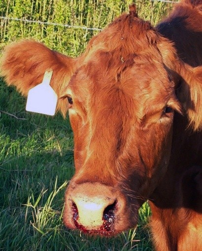
• This is a highly infectious viral disease of farm animals.
• This disease mostly manifests the lesions in the mouth, feet and mammary gland.
• Milk yield drops dramatically in milking animals, suckling calf usually die and pregnant animals may abort and infertility may ensure following abortion.
Causes
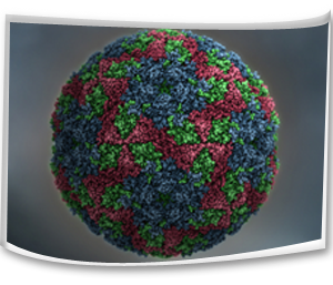
• It is caused by a virus Apthous of the family Picornaviridae.
• It has seven immunologically distinct serotypes namely O, A, C, Asia1, SAT1, SAT2, SAT3.
• The virus is quickly inactivated outside the pH range of 6.0 - 9.0 and by desiccation and temperature more than 56˙C, although virus may survive a considerable time when associated with animal protein such as in infected milk the virus will survive pasteurization at 72˙ C for 15 seconds.
• The virus is resistant to alcohol, ether and chloroform.
Mode of Transmission
• Generally by direct or indirect contacts between susceptible and infected animals.
• Through movement of clinically affected animals.
• Through inanimate vectors such as vehicles, fodders, utensils, equipments etc.,
• Through air. Infected animals have a large amount of aerosol virus in their exhaled air, which can infect other animals via the respiratory or oral routes. The virus can travel up to 60 km overland and 300 km by sea.
• All secretions and excretions from the infected animal such as saliva, faeces and urine. The virus may be present in milk and semen for up to 4 days before clinical signs appear.
• The disease has been transmitted to calves via infected milk.
• This virus can survive in dry fecal material for 14 days in summer, in slurry up to 6 months in winter, in urine for 39 days and on the soil between 3 (summer) and 28 days (winter).
• By consumption of infected meat and meat by-products, unprocessed and uncooked milk.
• Through animal handlers, visitors and physicians.
• Most of the animals remain as a carrier following recovery after infection. Carrier may transfer the virus from one animal to another. Carrier cattle may harbor the virus in the esophageal-pharyngeal fluid for 6-24 months.
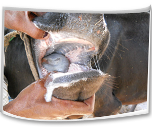
Symptoms
Clinical symptoms
• High fever up to 104-106˙F (41˙C) and anorexia.
• Profuse salivation (saliva hanging in long ropy strings up to the ground).
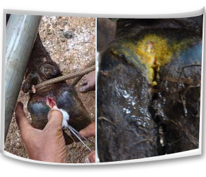
• Animal stamps its feet and wounds in the interdigital space of legs followed by lameness.
• Oral ulcers and lesions.
• Smacking of lips.
• Vesicles in the mammary gland.
Management Methods
Preventive measures
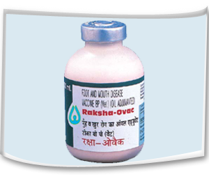
• Regular vaccination of farm animals, first dose at 3 months of age, followed by second dose at 30 days after first vaccination. Then repeated once in 6 months interval preferably during April- May.
• Vaccination of all the animals of an area/village is to be done at one time.
• Ring vaccination may be followed for control of disease outbreak and border vaccinations to protect disease free zones.
• Only vaccinated animals should be brought into the village from outside sources that too only 15-21 days following vaccination.
• No purchase of animals from disease prevailing areas.
• New animals should not be purchased until six months following outbreak.
• Unvaccinated animals should not be allowed to cattle fairs.
• Strict quarantine measures for newly purchased animals.
• A foot bath or truck bath may be made at the entrance of the village/farm.
• Always prefer to purchase / procure fodder from a place where FMD has not been recorded for a period of six months or so.

Suggested first aid
• Separation of affected animals from other animals.
• Mouth and feet of the affected animals should be washed with 1% potassium permanganate (KMnO4) antiseptic mouth wash3-4 times a day.
• Glycerin may be applied over the lesions.
• Antibiotic treatment and consultation should be made with qualified veterinary Doctor.
Control measures
Isolation and confinement of affected animals immediately after detection of clinical symptoms and restriction of animal movements.
Infected animals should not be allowed to graze in common grazing pasture.
Affected animals should not be allowed to drink water from ponds/streams/ rivers etc.
Diseased animals should not be allowed to roam about with other animals of the village.
Diseased animal handlers and attendants movements should be restricted to the other animal population / farms. If it is not practicable, people should scrub themselves and their belongings with soap and caustic soda.
In case of outbreaks, healthy animals should be attended first and then the affected ones. After attending the sick animals, persons should wash himself and his clothes with 4% sodium carbonate solution. Utensils used for collecting milk should be cleaned with 4% sodium carbonate solution.
Calves should not be allowed to suckle affected mothers and they should not be fed with milk from affected animals.
Mouth of the affected animals may be washed with antiseptic mouth wash. 1% potassium permanganate solution may be applied 3-4 times a day.
Feet of the affected animals may be washed with 2% copper sulphate solution. Antiseptic lotion and fly repellents are to be used to avoid infection and maggot formation on the wound.
Disinfection of floors, premises and all infected materials by using Sodium hydroxide (2%), sodium carbonate (4%) and citric acid (0.2%) is advisable.
Lime powder should be sprinkled around the animal houses.
Foot bath should be made at the entrance of the farm.

Anthrax
About the Diseases
Nature of disease
• It is an acute infectious disease of livestock that occurs throughout the world.
• This disease is also known as splenic fever due to the fact that there is extensive enlargement of the spleen(splenomegaly) due to this infection.
• Most of the food animals are affected with anthrax.
• No mammals have got absolute natural immunity against anthrax.
• The most susceptible animals are cattle and sheep.
• It is a zoonotic disease.
Causes
• The disease is caused by bacteria known as Bacillus anthracis.
• When the organisms are exposed to air (oxygen), spores are formed. The spores are never formed so long the organisms remain in the circulation. But when the organisms come out of the body, the spores are formed.
• The spores are very much resistant to cold, hot, chemicals and drying.
• The spores may remain viable in the soil for a considerable period of time and for ten years in the infected tissues and cultures.
• The soil can maintain the organisms in spore stage for years together without endangering the life of animals.
• The spores remain resistant to 100˙C for 5 minutes. But it will be destroyed at 100˙C for 10 minutes.
• The vegetative form of the bacteria can be killed at 60˙C for 30 minutes. In autoclave at 120˙C for 15 minutes all the vegetative forms can be killed.
• Commonly available chemicals cannot kill the spores.
• 5% NaOH can effectively destroy the spore contaminated objects.
Mode of Transmission
• The anthrax spores have got the ability to remain viable in the soil for a considerable period of time and thus remain as a continuous source of spread to the susceptible animals.
• The stream, rivers and flood may carry the spores from place to other and thus may spread the disease to the virgin soil.
• Carnivore animals may carry the infection to the distant places. Carnivores may contact the infection through ingestion of contaminated carcases.
• Flying birds may disseminate the infection from one place to the other.
• Various flies have been implicated as carrier of infection during the fly breeding seasons.
• Animals while graze in the infected pasture pick up the infection through ingestion or through breach in the oral mucosa or skin.
• The new area may be infected due to contaminated animal products such as bone meal, fertilizers, hide, hair, wool, grain or forage.
Symptoms
Clinical symptoms
• There is elevation of body temperature (104 to 108˙C).
• Animal refuses to eat and there is development of bloat.
• Animal is extremely depressed. Animal shows distressed breathing.
• Extreme dyspnoea leads to mouth breathing due to oxygen hunger.
• Sudden death within 48 hrs of illness of animal
• Following death there is oozing of blood from the natural orifices.
• Oedema may predominantly notice under the neck, brisket region, thorax, abdomen and flank.
• In per- acute form animals may be found dead without any premonitory signs.
Management Methods
Preventive measures
• Periodical and regular vaccination should be done.
• Strict quarantine measures in anthrax prone areas.
• Preventing the introduction of infected animals into disease free areas.
• Carcasses should not be opened as it may contaminate the pasture.
• Care should be taken to destroy the dead body by deep burial with quick lime.
• Persons handling the anthrax infected animals should adopt adequate sanitary measures.
• The adjacent areas of the dead and infected animals should be thoroughly disinfected by 3% per acetic acid or 10% caustic soda or 10% formaline.
• The fodder from infected pasture should be destroyed and not to be given to the other animals.
Suggested first aid
• The dead animal body should not be opened.
• Should have consultation with nearest qualified veterinary doctor.
• This disease should be brought under the notice of the regulatory officials in case of an outbreak.
• Care should be taken to destroy the dead body by deep burial with quick lime.
Zoonotic importance
• Anthrax is a zoonotic disease and thus has public health significance.
• Anthrax bacilli or spore may produce cutaneous abscess known as “hide porter’s disease”; pneumonia known as “wool sorter’s disease” or dysentery in man.
• Animal clinicians should take care while making blood smear from dead animals.
• Persons handling the anthrax infected animals should adopt adequate sanitary measures for their own safety.
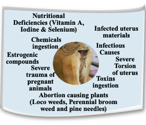
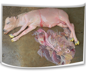
Abortion
About the Diseases
Nature of disease
• Expulsion of a dead or live recognizable size fetus at any stage of gestation (45-60 days onwards to parturition) is called as abortion.
• This may be due to various causes
Causes
• Infectious conditions such as Bovine Viral Diarrhea (BVD), Infectious Bovine Rhinotracheitis (IBRT), Leptospirosis, Brucellosis, Mycotic abortion, Actinomyces, Trichomoniasis, Campylobacteriosis, Listeriosis, Chlamydiosis, and Epizootic Bovine Abortion may cause abortion in cattle.
• Severe trauma, severe torsion of the uterus and twinning may cause abortion.
• Toxins can cause abortion in cows.
• Coumarins from rat poison and moldy sweet clover can cause abortion.
• Nitrates, chlorinated napthalenes, arsenics can cause abortion.
• Locoweeds, perennial broom weed and pine needles can cause abortion.
• Estrogenic compounds may cause abortion.
• Douching and infusion or Artificial insemination of the pregnant animals causes abortion.
• Vitamin A, iodine and selenium deficiencies can cause abortion.
Symptoms
Clinical symptoms
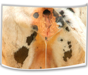
• Protrusion of fetal membranes or water bags from the vulva prior to expected date of calving.
• Discharges from the vulva.
• Spontaneous expulsion of a dead or live recognizable size fetus without full development.
• Anorexia, dull and depressed condition of animal.
Management Methods
Preventive measures
• Proper disposal of the aborted fetus and its membranes.
• Prevention of infected materials (uterine discharges, fetal membranes) ingestion.
• Always the animal shed should be clean.
• To prevent abortion due to Infectious Bovine Rhinotracheitis and Infectious Pustular Vulvovaginitis (IBR-IPV) pregnant cows at any stage of gestation should not be vaccinated with IBR-IPV vaccine. Vaccination can be carried out in heifers at 6-8 months of age.
• To prevent abortion due to listeriosis, feeding of poor quality silage with high pH should be prevented.
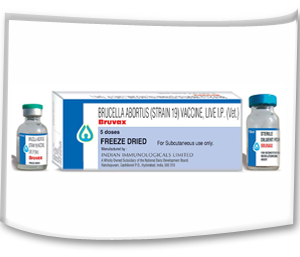
• To prevent abortion due to brucellosis, vaccination of calves from 3 to 7 months of age with strain 19 Brucella vaccine and bull calves should not be vaccinated.
• Breeding should be stopped at disease outbreak.
• Contact between animals should be kept at a minimum during outbreak.
• Diseased bulls should not be used for breeding.
• The semen which is used for Artificial Insemination should be free from any infectious agents.
• Pregnant animals should be protected from other animals attack.
• Pregnant animal sheds should not be slippery.
Actinomycosis
About the Diseases
Nature of disease
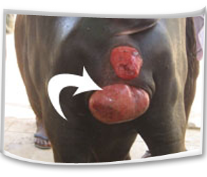
• This is a chronic infectious bacterial disease occurs in dairy animals.
• This causes swelling of lower jaw or around the mandibular region.
• This condition affects the feed intake of the animals.
• The disease is characterized by rarefying osteomyelitis of the bone of skull in cattle.
Causes
• Actinomycosis is caused by Actinomyces bovis.
• In addition to this organism, association of bacteria like Corynebacterium pyogenes and Staphylococcus are also seen.
Mode of Transmission
• Actinomycosis generally affects cattle between 2 to 5 years and it is a sporadic disease and animal to animal transmission occurs rarely.
• The organisms remain as resident population and may establish the infection through abrasion, injury or wounds.
• The abrasion of buccal mucosa induced by coarse feed or surface material while chewing may set up infection.
• Transmission of infection through dental alveoli at the time of eruption is noted.
• The alimentary canal of normal cattle may harbor A.bovis and from where the organisms may invade the subepithelial tissues through injury by surface object.
Symptoms
• The lesions appear initially as a hard, painless, circumscribed protuberance usually at the level of central molar teeth of the mandible or maxilla.
• The invasion damages the bony tissues and in some cattle, large granulomatous mass appear on the surface of the jaw followed by development of sinus tracts.
• Due to extensive involvement of the mandible and maxilla, the process of mastication is affected and thus there is impairment of digestion resulting to loss of general health.
• Abscess may extend and may produce sinus to the skin surface where from, the purulent discharges are drained.
• Examination of oral cavity may exhibit loose teeth or missing teeth.
• There is foul breath from the mouth known as halitosis.
• Loose teeth induce hypersalivation and dysphagia (difficulty in feeding).
• The adjacent bones may be affected in long standing cases.
• The adjacent lymph nodes are not affected and the disease does not spread through lymphatic channel.
 Management Methods Control measures
• There is no vaccine against this disease.
• Isolation of infected animals and their treatment are to be rendered.
• Removal of contaminated materials and disposal of animals with discharging foci may be made.
• This condition should be consulted with qualified veterinarian for antibiotic treatment.
Ephemeral fever
About the Diseases
Causes
• It is an arthropod transmitted viral disease of cattle and buffaloes.
• Among the cattle age group ranging from 6 months to2 years are more susceptible.
• The virus belonging to rhabdo virus group.
Mode of Transmission
• The disease is transmitted by sand fly.
• Mosquitoes like culex, culicoides have been suggested as a disease transmitter.
• Transmission does not occur by direct contact from animal to animal or via their discharges.
• Outbreak generally used to occur in summer season.
• The virus spread rapidly by wind and spread of the virus over three thousand miles within 5 months period has been reported.
• After infection the virus is found in the blood of cattle in about 5 days. The insects pick up the virus from the blood. Even 0.002 ml of blood may produce disease in susceptible hosts.
• The viruses never exist in the recovered animals and it is suggested that some fauna may be responsible for carryover of the virus from season to season.
• The disease can be transmitted by injection of whole blood.
Symptoms
Clinical symptoms
• The disease is preceded by a sharp rise of temperature ranging between 103-107˙F or more.
• There is shivering and muscle trembling.
• The affected cattle decline to move and if forced to move, they move with great difficulty with arched back condition.
• There is marked anorexia and appreciable reduction in milk yield.
• Salivation, nasal secretion and lacrymation are noticed.
• Muscle of the affected limb becomes stiff, hard and painful.
• The animal shows lameness to acute laminitis.
• Severely affected cattle may lie down with extended rigid hind limbs.
• Lameness may shift from one leg to other within few hours.
• The recumbent animals will also show suspended rumination, grinding of the teeth and signs of groaning.
Management Methods
Control measures
• Vector control is the important key to curb down this disease propagation.
• At present no commercial vaccine is available.
Parasitic Diseases of Cattle and Buffalo
A parasitic disease is an caused or transmitted by a. Many parasites do not cause diseases. Parasitic diseases can affect practically all living, including and. Some parasites like and can cause disease directly, but other organisms can cause disease by the that they produce.
Although organisms such as function as parasites, the usage of the term "parasitic disease" is usually more restricted. The three main types of organisms causing these conditions are (causing), Protozoa and helminths are usually endoparasites (usually living inside the body of the host), while ectoparasites usually live on the surface of the host.
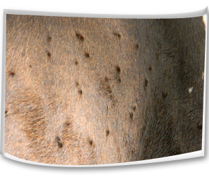
Anaplasmosis
About the Diseases
Nature of disease
• It is an important rickettsial infectious disease of ruminants.
• Exotic and crossbred cattle are highly susceptible.
Causes
• The disease is caused by Anaplasma marginale.
• The anaplasmas are intra-erythrocytic bodies.
• They can be destroyed by heating at 60˙C for 60 minutes.
Mode of Transmission
• The infection spreads through ticks.
• Besides tick, Tabanas spp., Stomoxys spp. And mosquitoes have been found to transmit the disease.
• Carrier animals like cattle and other wild ruminants play vital roles in the transmission of the disease.
• Mechanical transmission through dehorning, castration, vaccination, ear marking has been suggested.
• Transplacental transmission has been observed.
Symptoms
Clinical symptoms
• High rise of temperature,
• Loss of condition,
• Nasal discharge, lacrymation,
• Inappetance,
• Coughing, dry rales, moist rales,
• Rumen atony, dehydration, rough body coat, dyspnoea and muscle tremors.
• Enlargement of superficial lymphnodes,
• Grinding of teeth,
• Pale and icteric mucous membrane.
 Management Methods
Control measures
• This condition should be handled with qualified veterinary doctor.
• Strict control of insect population should be made by acaricidal spray or dips.
• Carrier animals should be isolated and disposed.
• Serological test of the herd should be made and the positive one should be brought under treatment.
• Prophylactic immunization against Anaplasmosis is done by preimunition, attenuated vaccine of ovine origin and inactivated vaccine of ovine and bovine origins.
Theileriosis
About the Diseases
Nature of disease
• Theileriosis is an important disease in exotic and cross bred dairy cattle
Causes
• The species of Theileria those affect cattle are T.annulata, T.parva and T.mutans.
• T.annulata is the most extensively distributed parasite and causes tropical Theileriosis.
Mode of Transmission
• The disease is mostly observed during summer and rainy seasons.
• This preponderance is attributed to higher activity of vector during this season (May to October). This is also influenced by stress due to hot and humid weathers.
• The disease is transmitted by the bite of the ticks of the genus Hyalomma.
• Erythrocytic forms of the parasite are ingested by the larval and nymphal stages of the tick along with blood, while feeding on an infected animal.
• The subsequent stages of the tick (nymph and/or adults) transmit the disease during their feeding on susceptible hosts (Transtadial transmission).
• The disease can also be transmitted mechanically by inoculation of infective blood and tissue suspension made from spleen, lymph node and liver of infected animals.
Symptoms
Clinical symptoms
• High rise of temperature (40.5˙C to 41.5˙C).
• Enlargement of regional superficial lymph nodes.
• Anorexia, restlessness and rough coat.
• Tense eye balls along with watery lacrymation.
• Laboured respiration, serous, nasal discharge and coughing.
• Depression and petechial haemorrhages on conjunctiva.
• Declination of fever and development of anaemia with high coloured urine in later stage.
Urticarial type skin lesions.
• Weakness, prostration and death.
 Management Methods
Control measures
• Tick population should be controlled.
Metabolic diseases of Cattle and Buffalo
About the Diseases
These are diseases of livestock caused by productivity practices when the body reserves on calcium, magnesium or energy cannot meet the metabolic needs. They are very important in places where high producing animals are required, e.g. in diary industry. In cattle, metabolic diseases include ketosis, milk fever, fat cow syndrome, and hypomagnesaemia. All these can produce an acute, temporary, but potentially fatal deficiency. Correcting the diet for cows during the period from late pregnancy to peak lactation is crucial in preventing these diseases. If these diseases occur frequently, it is essential to seek professional veterinary and nutritional advice.
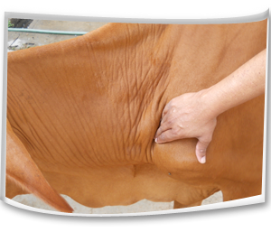
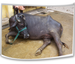
Downer cow syndrome
About the Diseases
Nature Of Diseases
• Affected animals remain bright and alert but are unable to stand.
• This is frequently met in exotic and cross bred dairy cows.
• Most commonly occurs immediately after parturition.
• Most commonly it is a complication of milk fever condition
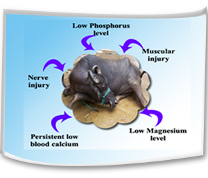
Causes
• Downer cows are unable to rise after two injections of calcium preparation indicating persistant hypocalcaemia.
• There is low phosphorus level.
• There may be of low blood potassium level. This is often seen in association with hypophosphataemia.
• A low level of blood magnesium has been incriminated as cause but it may develop along with low level of calcium.
• Muscular injury due to too much confinement in the byre, obesity, over feeding during dry period and too much compression of limbs.
• Following parturient paresis a cow may develop downer syndrome due to nerve injuries and over stretching of nerves or due to pressure on nerves while in recumbency.
• The damage of the heart muscle may be attributable to repeated dosing with calcium preparations in milk fever condition.
• Well fed highly obesed cows during later part of pregnancy very often suffer from a condition known as fat cow syndrome which predispose to downer condition.
 Symptoms
Clinical symptoms
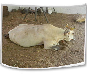
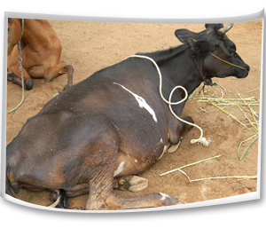
• Cow is inability to rise and remains in recumbent position.
• On stimulation the cow either makes little attempt or no attempt to rise or simply unable to rise even with treatment for milk fever.
• Cow remains bright and alert.
• Appetite, rumination, defecation and urination are usually normal.
• Temperature is usually normal but may turn towards sub-normal range in the terminal stage disease.
• The affected cow usually crawls around utilizing the forelimbs whereas hind limbs remain in flexed position. This type of stance is ascribed as "creeper cow"
• A downer cow which continues to remain down for more than 7 days ends fatally.
 Management Methods
Preventive measures
• We should provide most comfortable bedding prior to calving and in advanced stage of pregnancy. Sand is the ideal bedding material.
• Early detection and treatment of milk fever.
• Recently calved animals should be monitored at least 48 hours after parturition for the occurrence of milk fever signs.
• Recumbent animals should be treated as soon as possible and not delayed for more than 1 hour.
• Cow should not be mated with a heavy bull. The weight of the bull should be within the weight bearing capacity of the cow. Otherwise there is risk of paralysis and fracture of hip bones.
• The cow should be bred with a bull as per its size as a big calf in a small cow will invite dystokia problem leading to calving paralysis.
• Cow should not be made over fatty through too much feeding during advance pregnancy.
• Cow should be made to stand within a short time following parturition.
• Parenteral Vitamin D3 should be given in milk fever prone cow during pregnant period.
• Low calcium and high phosphorus diet should be given to stimulate parathyroid gland and thus to avoid hypocalcaemia.
• If possible cow of a dairy farm should be brought under metabolic profile test to pinpoint the deficit and to make good use of it.
 Control measures
• Recumbent animals should be treated as soon as possible and not delayed for more than 1 hour.
• Provision of comfortable bedding materials for recumbent animals. Arrangement of soft bed should be made.
• Attempt should be made to roll the cow from side to side to minimize the extent of ischemic necrosis.
• Lift the cow and frequent turning should be made. Cow should be turned at least at 3 hours interval.
• Attempt should be made to lift the cow on its fore legs by using body slings.
• Hip lifters may be used for lift the downer animals.
• With the help of body slings, the animals should be allowed to stand for 20-30 minutes and then lowered down. This should be repeated several times a day.
• Animal’s both fore and hind limbs should be massaged two times per day.
• Downer animals should be milked normally and the udder kept clean by washing with germicide soap before milking and post milking teat dips should be applied.
• Re-placement therapy with Calcium, Phosphorus, magnesium, Glucose containing preparations can be used parenterally by qualified veterinarian.
• Infective causes should be brought under antibiotic coverage.
• Physiotherapy by adopting muscle massage may be made to restore muscle activity of the limbs.
 Parturient paresis (Milk fever)
About the Diseases
Nature Of Diseases
• Parturient paresis is a metabolic disease occurring most commonly within 72 hours of parturition in adult females.
• It is frequently found in high yielding Jersey cows.
• Amongst cattle, mature and old cows are most commonly affected usually in the 5-10 year age group.
• High yielding dairy cows are mainly susceptible to the disease especially during their 3rd, 4th or 5th pregnancy or parturition.
• This is characterized by recumbency, muscle tremors and becomes normal after calcium borogluconate ingestion.
Causes
• A depression of the level of ionized calcium in tissue fluids.
• Excessive drainage of calcium in the milk just after parturition.
• Excessive loss of calcium in the colostrum beyond the capacity of absorption from the intestine and mobilization from the bones to replace.
• An impairment of absorption of calcium from the intestine at parturition.
• Deficiency of Vit. D and less acidic pH in gut.
• The mobilization of calcium from storage in the skeleton may not be sufficiently rapid to maintain normal serum level.
• Sometimes milk fever is associated with hypocalcaemia, hypophosphataemia and hypomagnesaemia.
• There is a special susceptibility if the animal is subjected to any stress. Forced exercise, long distance transport, sudden deprivation of food and grazing on oxalate containing plants or green cereal crops may precipitate the condition.
• Non –parturient Milk Fever occurs in any situation which may impose severe stress or starvation of feed deprivation may be the factors which may ultimately produce a sudden shift in calcium balance leading to hypocalcaemia.
Symptoms
Clinical symptoms
• There is muscle weakness and flaccidity of muscles.
• Tremor is found in the eye and muzzle.
• Animal may rest on sternum.
• There is diminished consciousness and the animal may show drowsy condition.
• There is lateral kink in the neck or head may rest on the flank (‘S’ shaped posture).
• The skin and the extremities remain cold. Temperature is generally subnormal.
• Muzzle becomes dry.
• Mucous membrane of the eye also turns dry with dilated pupil.
• Eyes are unable to blink.
• There is relaxation of anus.
• Venous pressure is low as such it may difficult to raise the jugular vein.
• There is atony of the rumen leading to constipation.
• The animal may be in lateral placement.
• There is complete flaccidity for which animal cannot sit up.
• Due to recumbency bloat is evident.
• There may be Anuria and oliguria due to paresis of muscles
 Management Methods
Suggested first aid
• Treat the animals as early as possible before the cow become recumbent.
• Bring the animal to sternal recumbency until treatment is available.
• Bring the recumbent cows from slippery floors to non-slippery areas.
• If the animals are lying in the open ground, erect a temporary shelter to protect from sun light.
• Immediately after evidence of milk fever signs and recumbency, consult with qualified veterinarian for calcium administration.
Prevention Method
• A diet containing less than 20 gm of calcium per day should be fed during the last two weeks prior to calving to prevent milk fever.
• Avoid excess calcium intake during the dry period. Diet containing less than 80 to 100gm/ day of calcium throughout the dry period may be fed to prevent milk fever.
• Phosphorus intake of less than 35 gm/ day may be the standard level for the prevention of milk fever.
• High phosphorus and low calcium level during the last month of pregnancy may be maintained to prevent milk fever (Ca: P= 1:3.3).
• Diets containing higher level of chloride and sulphur - anions relative to sodium and potassium cations may prevent occurrence of the diseases. Excess anions help in the absorption of calcium.
• Administration of ammonium chloride 3 weeks before parturition may be a useful method of prevention.
• Use of Vitamin D3 and its metabolites has been an effective approach to prevent milk fever. A single intramuscular injection @ 10 million unit 3-8 days prior to parturition may be made. A dose of 1 million units for every 45 kg body weight would be a rational preventive approach.
• Oral calcium gel dosing (50% calcium chloride) has been suggested at prior to calving, at calving, 12 hours post calving and 24 hours of post calving to prevent milk fever.
• Addition of lime stone water in the drinking water prior to parturition will prevent this condition.
• Avoid over fattening in the pre-partum period.
• Avoid stresses at the time of parturition.
• Provide a clean, well- bedded box stall for calving.
• Make frequent observation of cows prone to milk fever from 48 hours after parturition for signs of milk fever and treat them promptly if any signs are exhibited.

 Control Method
• Calcium borogluconate-25% 500 ml slow Intravenous injection 10-20 drops/ minute.
• Compounds containing cal-Mg-Boro-gluconate-200 to 350ml I/v followed by S/c for rest of dose.
 Obstetrics and Gynecological conditions of Cattle and Buffalo
Anoestrum
About the Diseases
Nature Of Diseases
• This is a metabolic disorder of dairy animals.
• This manifest digestive disturbance.
Causes
• Failure to detect / observe oestrus signs.
• Suboestrus, weak or silent oestrus.
• A low plane of nutrition, lack of energy and protein, deficiency of minerals namely P, Co, Fe, Cu, I, Mn and Vitamin A.
• Failure to recognize that an animal is pregnant.
• Anoestrus due to persistent corpus luteum, conditions associated with uterine pathology such as pyometra, mummified foetus, foetal maceration, mucometra and hydrometra and
• Insufficient hormonal stimuli.
• Senility, chronic debilitating disease like JD, TB etc., seasonal and environmental influences and heavy lactation (negative energy balance) may predispose anoestrus.
• Closely confined dark stables, lack of exercise combined with nutritive factors.
• In suckling animals prolactin may reduces the ovarian sensitivity
Symptoms
Clinical symptoms
• Absence of oestrus signs
Management Methods
Prevention Method
• Unobserved oestrum may be due to managerial deficiencies and short period of oestrus. The dairy animals should be observed for heat signs at least three times a day.
• Wall charts, breeding wheels, herd monitors and individual cow records may be used for identify the oestrus.
• Teaser bulls (vasectomized or by applying apron) are useful in identifying heat in large number of animals especially buffalo cows.
• Provision of adequate lighting to improve oestrus detection.
• Silent / weak / Suboestrus are most common in buffalo cows and common in post partum period. In this cyclical changes in the genital organs occurs but the signs of heat are not exhibited or not observed. This requires rectal examination by qualified veterinary doctor.
• Extra feeding of a concentrate mixture or grains like maize, Cholam, kambu. Etc., and at least small amount of green fodder along with other roughages.
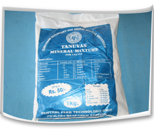
• TANUVAS Mineral mixture may be supplemented.
• After breeding the animals should be checked for pregnancy within 45-60 days by qualified veterinary doctor.
• Uterine pathology and hormonal stimuli should be handled by qualified veterinary doctor.
 Repeat breeder
About the Diseases
Nature Of Diseases
• Animals come to heat and not conceived after three successive inseminations are called repeat breeder.
• This may be due to various reasons
Causes
• Due to deficient Luteinizing Hormone release, delayed ovulation or failure of ovulation may leads to fertilization failure.
• Defective ovum or ageing of ovum may leads to fertilization failure.
• Inability of the sperm to fertilize a viable ovum.
• Inability of gametes to reach one another.
• The organisms Trichomonas fetus, Campylobacter fetus, Brucella abortus and IBR-IPV which may cause early embryonic death.
• Deficiency of Selenium and Vitamin E may cause early embryonic death.
• Long period of feeding estrogenic forages may affect the embryo survival.
• Environmental stress during first week after breeding may lead to early embryonic death.
Symptoms
Clinical symptoms
• Animal will not conceive even after three successive inseminations.
Management Methods
Preventive and Control measures
• Bring the animal into positive nutritive balance.
• Mineral mixture supplementation should be done to breeding animals.
• Do Artificial Insemination twice at each oestrus preferably at 12 or 24 hrs intervals.
• Skipping of AI and intrauterine infusions may be considered for uterine pathology.
• Diseased bulls should not be allowed for breeding.
• By avoiding diseased breeding bulls the pathogenic organisms causing abortion may be controlled.
Endometritis
About the Diseases
Nature Of Diseases
• Endometritis is a localized inflammation of the uterine lining, associated with chronic postpartum infection of the uterus with pathogenic bacteria.
Causes
• The causal organisms usually reach the uterus from the vagina at coitus, insemination, parturition or postpartum.
• The great majority of cows suffer from bacterial contamination of the uterus after calving.
• In cows that develop endometritis, the bacterial flora is not eliminated from the uterus, causing the endometrium to become inflamed.
• The factors associated with the development of endometritis are, retained fetal membranes, abortion, induced calving, multiple births, dystokia and bacterial loading.
Symptoms
Clinical symptoms
• The presence of a white or whitish – yellow mucopurulent vaginal discharge in the post partum cow.
• The volume of discharge is variable, but frequently increases at the time of oestrus when the cervix dilates and there is copious vaginal mucus.
• The cows rarely show any signs of systemic illness, although in a few cases milk yield and appetite may be slightly reduced.
• Rectal palpation frequently shows a poorly involved uterus which has a doughy feel.
Management Methods
Control measures
• A wide range of antibiotics, hormones, antiseptics and immunomodulators have been used as treatments for endometritis.
• This condition should be handled with qualified veterinary doctor.
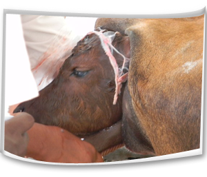
Dystocia
About the Diseases
Nature Of Diseases
• The first or the second stages of parturition is markedly prolonged, becomes difficult or impossible for the dam to deliver the foetus without artificial aid.
• Dystocia means difficulty in birth.
• The overall incidence of dystocia varies with the species and with breeds within the species.
• The bovine species is most often affected.
• Dystocia is common in primipara than in pleuripara.
• Heavier male calves, twin pregnancy in cattle and low litter size in multiparous species, increase the incidence.
• Pregnancies that terminate early are conducive to dystocia through the medium of uterine inertia and fetal malposture.
• Prolonged gestation causing fetal oversize, close confinement, overfeeding, gross underfeeding and too early breeding increases the incidence.
Causes
• Twining, hydro amnion and foetal anasarca
• Improper nutrition of the growing heifers was the most important factor in retarding body and pelvic growth.
• Small pelvis, under developed juvenile genital tract, and lack of strength to expel the foetus.
• Breeding a poorly grown, underfed female that may be old enough to breed, but the body growth has been greatly retarded due to poor nutrition, parasitisms or diseases.
• High feeding levels favours excessive deposition of fat in the pelvic region predisposing to difficult parturition, especially in heifer.
• High feeding levels favours the development of a larger fetus (especially high feeding during the last third of pregnancy) leads to difficulty in giving birth.
• Close confinement of pregnant animals without exercise, are prone to torsion of uterus and uterine inertia.
• Any infection or disease affecting the pregnant uterus and its contents may cause dystocia.
• Dystocia may be due to Expulsive forces (Expulsive defect), adequacy of the birth canal (Constriction) and size and disposition of fetus (Over size and faulty disposition).
Symptoms
Clinical symptoms
• Difficulty in giving birth.
• Calf limbs or face protrude from the vulva.
• Animal attempts strong forces and unable delivers calf
Management Methods
Control measures
• It should be handled with qualified veterinary doctor.
• It has been suggested that dairy heifers may be bred by size or weight rather than by age.
• During parturition all animals should be watched closely, if possible, so that prompt aid may be given if parturition is not normal.
• To help control infections that predispose to uterine disease and foetal death, both the sire and dam should be free of infection at the time of service.
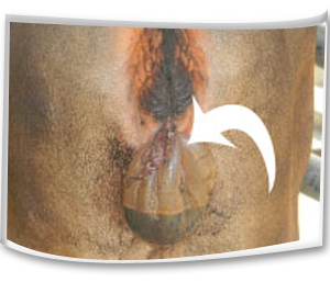
Total Uterine Prolapse
About the Diseases
Nature Of Diseases
• This is expulsion of uterine mass through the vagina.
• It is a common complication of third stage of labour.
• It is more common in pluriparous than primiparous animals.
• This condition is most commonly seen in cow than other animals.
Causes
• The flaccid atonic uterus.
• Violent or strong tenesmus during or after parturition.
• Retention of placenta at the ovarian pole of the uterine horn.
• Excessive relaxation of the pelvic and perineal region.
• Commonly seen in confined or stabled cattle especially during winter months.
• Forced extraction of the fetus.
• Over distension of the abdomen or excessive amounts of loose pelvic fat favour the condition by increasing the intra-pelvic pressure.
• Due to intra abdominal pressure.
• Delayed contraction of the uterus.
• Poorly grown, thin debilitated heifers and low plan of nutrition.
• During last 2-3 months of gestation, when large amounts of oestrogenic hormone being secreted by the placenta known as hyper estrogenism
Symptoms
Clinical symptoms
• A mild protrusion of the vaginal mucous membrane through the vulval lips when the animal lies down.
• In standing posture, the prolapsed mass hanging upto the hock joint.
• The fetal membranes and /or mucus membrane of the uterus is exposed.
• The mass is usually covered with feces, straw, dirt or blood clots.
• Uterus is enlarged and edematous especially in delayed cases (4-6 hrs).
• The cervix is usually present at the vulva.
• The non gravid horn is held inside the peritoneal surfaces of the prolapsed gravid horn and does not evert.
• Stress, restlessness, pain, anxiety, increased respiratory rate may be noticed.
• Internal hemorrhage due to rupture of one of the uterine vessels, shock, incarceration of the intestinal mass and death.
• Pale mucus membrane, expiratory grunt and prostration with severe depression and inability to rise indicated serious complications.
Management Methods
Suggested first aid
• The prolapsed animal should be separated from other animals.
• The mass should be protected from contamination from out sources.
• The mass should be covered with wet clean clothes until treatment.
• It needs careful insertion of protruded uterine mass into the body and suturing the in the vulval lips so this condition should be handled with qualified veterinary doctor.
Torsion of Uterus
About the Diseases
Nature Of Diseases
• Uterine torsion is defined as the twisting or revolution of the gravid uterus on its long axis and is common in cows and buffaloes.
• High incidence among pluriparous than primiparous
Causes
• Frequent lying down and getting up may predispose to uterine torsion.
• Lack of fetal fluids and violent falling or rolling and sudden movements may predispose to torsion.
• Close confinement of pregnant animals for a longer period may favour occurrence of uterine torsion.
• A lack of tone of the pregnant uterus comprising lack of fluids, flaccid uterine walls, a small non gravid horn, a long flaccid mesometrium favours uterine torsion.
• A deep capacious abdomen predispose to uterine torsion and especially in buffaloes because of the wallowing habit.
• Exiting causes such as horn thrust or butting by the neighboring animals, violent movements during grazing, rolling due to tympany and colic may cause torsion.
• Transportation of pregnant animals from one place to another in rail or by road cause violent and irregular movements creating anxiety and predispose to torsion of uterus.
Symptoms
Clinical symptoms
• The signs of abdominal pain, anorexia, constipation, lack of ruminations, restlessness or colic symptoms, teeth grinding, treading and tail switching may be observed.
• Displacement of dorsal commissure.
• Tucked up udder and
• Vulval edema.
Management Methods
Prevention Method
• Pregnant animals should be housed separately and extra care should be given in handling the animals.
• Rolling, jumping, frequent lie down and falling of pregnant animals should be avoided.
• Transportation of pregnant animals should be avoided.
• Attack and fight with other animals should be avoided.
Control Method
• This condition must be handled with qualified veterinary doctor.
• Rotation/ rolling of the dam may be performed with the help of veterinary doctor.
• In complicated cases cesarean or Laparo- hysterotomy may be recommended.
Retained Fetal Membranes
About the Diseases
Nature Of Diseases
• This is one of the most common conditions occurring in animals following parturition.
• This is failure of the villi of the fetal cotyledon to detach from the maternal crypts of the caruncles and retained longer than normal time limits.
• If the placenta is retained longer than 8-12 hours the condition is considered as pathological.
Causes
• Infections of uterus during gestation may be a cause for retained placental membranes.
• Uterine inertia due to hormonal imbalance such as low level of oxytocin.
• Deficiency of Vitamin A and iodine causes retained placenta.
• The hormone progesterone and excess of cortisol in late gestation may cause the retension of fetal membranes.
• The disease conditions causing uterine inertia or atony results in a higher incidence of retention of foetal membranes.
• Close confinement and lack of exercise highly prone to retained placenta.
Symptoms
Clinical symptoms
• A portion of the fetal membranes hang from the vulva 12 hours or more even after the expulsion of the foetus.
• Anorexia and depression may develop.
• A fetid odour develops since the placenta begins to macerate after 24 hours of foetal expulsion.
Management Methods
Control Method
• It should be handled with qualified veterinary doctor by manual removal.
• Before arriving veterinarian, the hanging foetal membranes should be protect from dogs or other animals.
• The protruding membranes should be tied in a knot to prevent them touching the hocks.
• Manual removal can be attempted in 24- 48 hours after parturition.
• Manual removal after 48 hours is not advisable because cervix was closed.
• Manual removal of placenta is contraindicated in cows with elevated temperature and also with vaginitis and vulvitis.
• After removing the fetal membranes, tetanus toxoid injection is recommended to prevent tetanus infection.
Bloat
About the Diseases
Nature Of Diseases
Prevention of coalescence of the small gas bubbles and entrapment of the normal gases of fermentation.
Production of stable foam.
Frothiness of ruminal contents.
Lush, young pastures and leaves containing high concentration of soluble protein and dominated legume plants particularly alfalfa, red and white clovers and occurs with grazing of young green cereal crops, rape, turnips and legume vegetable crops.
Feeding of high quality hay.
Feeding of high grain diet.
Feeding of the finely ground feed.
Physical obstruction to eructation occurs in esophageal obstruction caused by a foreign body, pressure outside the esophagus and obstruction of cardia.
Symptoms
Clinical symptoms
Obvious distention of the rumen and entire abdomen.
Discomfort with the animal may stand and lying down frequently, kicking at its abdomen and rolling.
Sudden death with distended abdomen.
Dyspnea and grunting accompanied by mouth breathing
Protrusion of the tongue and extension of the head.
Management Methods
Suggested first aid
The passage of a stomach tube or trocarization to release large quantities of gas.
An incision of about 10-20 cm in length over the left paralumbar fossa through the skin, abdominal musculature and directly into the rumen.
A stick is tied in the mouth like a bit to promote the production of excessive saliva.
Administration of antifoaming agents such as vegetable oils (peanut, corn, soybean) and mineral oils (paraffin) at doses of 80-250 ml.
Prevention and Control measures
The pasture should be free from leguminous fodders and bloat producing plants.
Feeding hay before turning cattle on pasture.
Maintaining grass dominance in the sward or using strip grazing to restrict intake.
Allowing animals on well grown mature pastures than immature or rapidly growing pastures.
Grass- legume mixture with a legume content of 50% is suggested as the maximum bloat safe level.
Prevention of high energy and high protein supplement.
Drenching of 60-120 ml of antifoaming agents twice daily (at milking times).
Feedlot rations should contain at least 10-15% cut or chopped roughage mixed into the complete feed. Preferably the roughage should be a cereal, grain straw, grass hay.
Grains should be rolled or cracked, not finely ground.
Pelleted rations made from finely ground grain should be avoided.
Enteritis
About the Diseases
Nature Of Diseases
• Inflammation of the intestinal mucosa resulting in diarrhea, dysentery, abdominal pain and varying degrees of dehydration and acid- base imbalance.
Causes
• The enteropathogens like bacteria, viruses, fungi, protozoa and helminths, chemical and toxins.
Symptoms
Clinical symptoms
• Diarrhea
• Dehydration, abdominal pain, septicemia and toxemia with fever.
• Feces are soft or fluid in consistency and unpleasant odor.
• Contain blood, mucus/ foreign materials like sand.
• Color of feces is pale yellow and sometimes frank blood.
• Distribution of the feces on animal’s perineum.
Management Methods
Preventive measures
• Ensure adequate non specific resistance by adequate colostrums intake.
• Vaccinate for those diseases for which there is an effective vaccine.
Control measures
• Reduce infection pressure.
• Minimize Managemental and environmental stressors.
Thelitis
About the Diseases
Nature Of Diseases
• This condition is inflammation of teat due to entry of pathogens into teat.
• It most commonly occurs in milch animals.
• It is due to unhygienic measures and environment.
• This causes no change in colour and consistency of milk.
• Untreated teat results in complete destruction of teat.
Symptoms
Clinical symptoms
• Affected teat initially shows reddening and swelling of teat.
• Infection progress leads to inflammation of teat.
• Decreased milk production.
• Severe infection leads to destruction of affected teat.
Management Methods
Prevention and control measures
• The animal’s environment should be clean and hygienic.
• The floor of the milch animal should be periodically cleaned with antiseptic solution.
• The milker’s hand should be clean before each milking.
• The dipping of teat after each milking may be effective in preventing entry of pathogens into teat.
• Affected animals should be given with earlier treatment to avoid destruction of teat.
• Affected animal’s teat should be treated with qualified veterinary doctor.
Traumatic Reticulo Peritonitis (TRP)
About the Diseases
Nature Of Diseases
• This is a disease condition commonly occurring in dairy animals by ingestion of hard objectives like nail, wire and iron materials through feed.
• These hard objectives after ingestion enter into the stomach and reach the reticulum and pierce the peritoneum and enter into the heart.
• The pregnant animals mostly affected than non pregnant animals.
• Most of this condition results in guarded prognosis.
Causes
• Caused by the ingestion and migration of a foreign body in the reticulum.
• The feed with hard objectives like nail, wires, hair pins, stitching needles and other piercing needles.
• Allowing the animals in pastures mixed with like these hard objectives.
Symptoms
Clinical symptoms
• Fever
• Anorexia
• An arched stance with abducted elbows
• Muffled heart sounds
• Jugular pulses
• Brisket edema
• Decreased milk production
Management Methods
Diagnostic tests
• Positive stasis test in affected animal- Stagnation of blood on either side of point of application of pressure.
• Negative stasis test in Normal Animal-Stagnation of blood on one side of point of application of pressure.
• Animal movement test- Affected animals will reluctant to move in slopes from up to down and move very slowly with difficulty in walk
Prevention and Control measures
• Feed is the main root cause and it should be free from any wires, nails and metallic objects.
• Animal houses and surroundings should be free from wires, nails, metallic objects and hardware materials.
• Cattle should be kept away from construction sites.
• Crop fields should be monitored for metal debris.
• Processed feed passed over magnets to recover any magnetic foreign bodies prior to being fed to cattle.
• In early stages, it may be consulted and treated by qualified veterinarian.
• In advanced stage, prognosis is guarded.