 |
||||||||||||||||||
|
||||||||||||||||||
| PARASITIC DISEASES | ||||||||||||||||||
| Back | ||||||||||||||||||
|
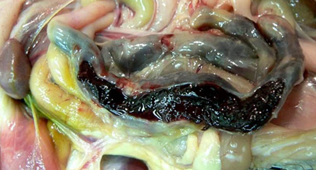
Coccidiosis (Eimeria spp.) Cause: Protozoan parasite of the intestinal tract of poultry. Most coccidia affecting chickens are in genus Eimeria (up to 9 species). Clinical signs: Pale combs and wattles from blood loss in gut. Ruffled feathers, depression, blood in droppings, shivering, decreased egg production by birds at reproductive age. Mortality rate may increase, particularly in caged birds. Gross Lesions Chickens: Lesions vary depending on what species of Eimeria are involved. Eimeriaacervulina: white “tiger-striping” of upper small intestine (duodenum). Eimerianecatrix: Mid and distal small intestines (jejunum and ileum), white spots (large schizonts) in mucosa with mucus and blood in lumen. Eimeriatenella: Hemorrhage in colon and cecum progressing to cores of coagulated blood in lumen of cecum. Eimeria maxima: Orange, mucoid material in lumen of jejunum. Eimeriabrunetti: Mucus and blood in ileum and colon, occasionally scores of blood in cecum. Diagnosis: Diagnosis is made by observing gross lesions and confirming presence of intestinal oocysts by histopathology. Alternatively, segments of infected intestine can be opened and the mucosa gently scraped with a glass coverslip. Place the coverslip on a glass microscope slide and identify the oocystscytologically Prevention and control: Coccidia can be attacked at three stages:
Blackhead:
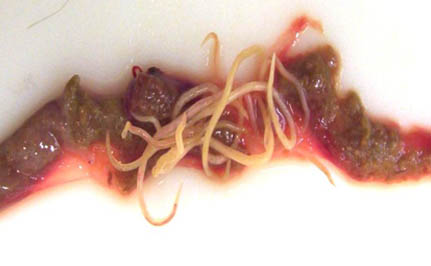
Trichomoniasis is caused by Trichomonasgallinae. It is a disease of the upper digestive tract. It has been found in pigeons, doves and kites but may invade hens and turkeys if they drink infected water or eat infected feed. Affected pigeons will go off-feed, appear ruffled, become emaciated and die, with a green yellow fluid dripping from the beak. Roundworms (Ascariasis) Cause: Ascaridiagallia nematode residing in the upper small intestine of chickens and turkeys Young birds less than 3 months are most susceptible to intestinal damage and light-weight breeds (e.g., White Leghorns) are more susceptible to infection. Infection can occasionally occur in caged layers that have been exposed to contaminated flies. Direct Life Cycle (no intermediate host). About 30 days to complete cycle. Clinical signs:Depression, weight loss, diarrhea, decreased growth, lowered egg production in heavy infection. Gross Lesions Worms in small intestine can occasionally cause death by blockage/impaction. Reddened intestinal mucosa (enteritis), thin bird, atrophy of breast muscle, and decreased body fat. Ascarids can migrate to oviduct and penetrate egg prior to shell formation. Diagnosis: Necropsy: identification of worms that are large, yellow-white, and 5-11 cm long. Fecal flotation (eggs float in zinc sulfate or sodium nitrate solution). Prevention/Treatment:Confinement and cage rearing has reduced problems with most intestinal parasites. Deep litter (4-6 inches of wood shavings) to reduce exposure to parasite eggs. Proper clean up between flocks. Treat pullets before they are transferred to laying house. Treatment: Piperazine.
Other Intestinal Parasites Species affected: All birds. Cause: Roundworms (Ascarids), Hairworms (Capillaria), Cecal worms (Heterakis), Tapeworms (Cestodes). Signs and lesions: Unthriftiness, stunted growth, emaciation, enteritis, anemia, decreased egg production. Prevention and control: Mainly relies on vaccination. Three types of vaccines are used:
|
||||||||||||||||||
|
|
||||||||||||||||||
| Back | ||||||||||||||||||
|
||||||||||||||||||
| Scroll | ||||||||||||||||||
| Division of Veterinary and Animal Husbandry Extension Education Faculty of Veterinary Sciences and Animal Husbandry, R.S. Pura, SKUAST Jammu | ||||||||||||||||||

